This focus idea is explored through:
Contrasting student and scientific views
Student everyday experiences
Younger primary students may have little knowledge about internal bodily organs. They tend to think the contents of the body are what they have seen being put into or coming out of it, such as food and blood. Their experiences with everyday cuts, scratches and bruises seem to reinforce a view that blood is below the surface of the skin, filling the spaces inside the body (like a bag of blood).
Older children are more likely to be able to list a large number of organs but may not fully understand the function or interconnected nature of these. For example, students at these levels may realise that the heart is a pump but not realise that the blood returns to the heart, or they may believe that the brain helps the body parts but not always realise that the body helps the brain.
Research: Fleer & Hardy (1996), Gellert (1962), Carey (1985)
Scientific view
 To survive and reproduce, the human body relies on major internal body organs to perform certain vital functions. When two or more organs along with their associated structures work together they become component parts of a body system.
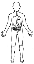
Some of the easily recognisable internal organs and their associated functions are:
The brain
The brain is the control centre of the nervous system and is located within the skull. Its functions include muscle control and coordination, sensory reception and integration, speech production, memory storage, and the elaboration of thought and emotion.
The lungs
The lungs are two sponge-like, cone-shaped structures that fill most of the chest cavity. Their essential function is to provide oxygen from inhaled air to the bloodstream and to exhale carbon dioxide.
The liver
The liver lies on the right side of the abdominal cavity beneath the diaphragm. Its main function is to process the contents of the blood to ensure composition remains the same. This process involves breaking down fats, producing urea, filtering harmful substances and maintaining a proper level of glucose in the blood.
The bladder
The bladder is a muscular organ located in the pelvic cavity. It stretches to store urine and contracts to release urine.
The kidneys
The kidneys are two bean-shaped organs located at the back of the abdominal cavity, one on each side of the spinal column. Their function is to maintain the body’s chemical balance by excreting waste products and excess fluid in the form of urine.
The heart
The heart is a hollow, muscular organ that pumps blood through the blood vessels by repeated, rhythmic contractions.
The stomach
The stomach is a muscular, elastic, pear-shaped bag, lying crosswise in the abdominal cavity beneath the diaphragm. Its main purpose is digestion of food through production of gastric juices which break down, mix and churn the food into a thin liquid.
The intestines
The intestines are located between the stomach and the anus and are divided into two major sections: the small intestine and the large intestine. The function of the small intestine is to absorb most ingested food. The large intestine is responsible for absorption of water and excretion of solid waste material.
Critical teaching ideas
- Humans may look different but inside they share identical component parts.
- The human body contains major internal organs or body parts which can be easily identified. These organs differ in size, shape, location and function.
- Each organ has a specific role which contributes to the overall wellbeing of the human body.
- A group of organs whose jobs are closely related are often referred to as a system.
 Explore the relationships between ideas about internal body organs in the
Concept Development Maps (Cell Functions).
Explore the relationships between ideas about internal body organs in the
Concept Development Maps (Cell Functions).
Building students’ understanding of internal body organs, how these are linked and why they work together as systems is a complex process. A useful starting point is to identify students’ existing ideas and understandings about what is inside the body. Using everyday experiences to draw out these ideas is always powerful, such as recalling visits to the doctor, medical operations/procedures, injuries, medical imaging/scans, posters and advertising images.
It is useful to explore what internal organs look like and where they are located in order to understand the specific function of each and how each contributes to keeping the body alive and well. Teaching experiences should begin to encourage students to consider how organs work together, i.e. how the work of one organ is similar or contributes to the functioning of another. This idea leads to the more complex idea that body parts form connected systems that contribute to the functioning of the body as a whole.
Teaching activities
Bring out students’ existing ideas
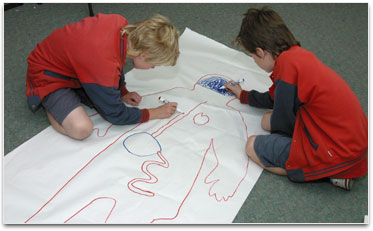
Encourage students to work in small groups to create a common drawing of what they know about the inside of the human body. Consider providing each group with an outline of a human body or have students trace around a group member lying on a large sheet of paper. Ensure students consider the location, size and shape of body parts in their drawings. Have students include labels naming each internal part and consider getting the groups to research information about each organ.
Share intellectual control
 Provide each student group with at least three strips of paper. Have each group list three questions that arose as they were completing their drawings, focusing on things they realised they didn’t know.
Display body drawings and discuss similarities and differences between each group’s representations. Display the questions and add further questions to the list as they arise from these discussions and observations. As a class, complete a bundling activity sorting the questions. Students should discuss what groupings to use.
These questions can then inform planning of further investigations. Revisit these questions at the end of each session and respond where appropriate with new information.
Clarify and consolidate ideas for/by communication to others
By using a jigsaw strategy, students move from ‘home’ groups to ‘expert’ groups, and then back to ‘home’ groups to collect and share more detailed information about internal body organs. Working in ‘expert’ groups, students research a specific internal organ of the human body. As a result of the experiences provided in this ‘expert’ group, each team member must be able to explain where the organ is located, describe/represent the observable features of the organ and explain what the organ does for the human body and why it is important.
When working in expert groups it may be useful for students to:
- access useful websites, science texts and visual images
- explore scientific models
- examine animal organs (following regulated health protocols)
- construct simple models with materials like plasticine
- complete simple investigations which demonstrate key features and functions of the organ they are investigating.
Each member then returns to the ‘home’ group and shares their expertise, ‘teaching’ their colleagues this new information. Following expert presentations the ‘home’ group returns to their original body drawing and adds new information. These changing body displays become an integral part of the ongoing investigation and demonstrate a dynamic, changing display.
Promote reflection on and clarification of existing ideas
To assist students to construct richer personal meanings for ideas and concepts related to internal body organs encourage them to complete sentence stems. Sentence stems are incomplete statements designed to provide a structure for insights and observations. Some examples are: ‘The liver is…’ ‘The liver can…’ ‘A heart has…’ ‘The lungs can…’
Encourage students to write short stories about one of the organs they have investigated, with a focus of ‘a day in the life’ of that organ.
Challenge some existing ideas and focus attention on hitherto overlooked detail
When students are familiar with a variety of major internal organs, provide activities that encourage students to consider how one organ is similar to or contributes to the work of another organ. Ask students questions such as, ‘How is this like/not like a similar organ?’ This encourages students to consider similarities and differences which they may normally overlook. Other questions might include:
- How are the stomach and bladder alike? How do they differ?
- How are the kidneys and the liver alike? How do they differ?
- How is the large intestine like a tea strainer?
- How is the heart not like a bike pump but more like an air mattress foot pump?
Identify diseases and conditions commonly associated with each major organ, such as heart attack, asthma, stomach ulcer, etc. Discuss contributing factors to these, such as inherited conditions. Explore how medicine has developed effective treatments for many of these, for example transplants, mechanical hearts and medication.
Further resources
Science related interactive learning objects can be found on the
FUSE Teacher Resources page.
To access the interactive learning object below, teachers must login to FUSE and search by Learning Resource ID:
-
Human Body – students work through a series of four learning objects about the structure and function of the human body. They compare humans with other animals to explore adaptation to environments.
Learning Resource ID: K6MKAS